
9 Best Exercises for SI Joint pain
Introduction
A common condition that affects the region where the spine and pelvis meet is sacroiliac (SI) joint pain, which frequently results in discomfort in the hips, buttocks, or lower back. Joint inflammation, instability, or changed biomechanics caused by extended sitting, bad posture, pregnancy, or repeated strain can all be the cause of this discomfort.
By specifically strengthening and extending the surrounding muscles, especially the core, glutes, and lower back, exercise can restore stability, increase mobility, and lessen discomfort in SI joint dysfunction.
By improving postural alignment and movement control, a well-organized program not only reduces present discomfort but also supports long-term joint health. To prevent irritating the joint, workouts should be chosen according to each person’s demands and executed correctly.
Anatomy of the Sacroiliac Joint
- The Articular surface: The sacrum of the vertebral column and the ilium of the pelvis articulate at the sacroiliac joint. A stable joint is created when the irregularly shaped articular surfaces connect. They have hyaline cartilage lining them. The joint cavity may be lost as a result of the sacroiliac joints becoming fully fused in later age.
- The joint capsule: One type of synovial joint is the sacroiliac joint. It is surrounded by a synovial membrane-lined fibrous joint capsule.
- The ligaments around the sacroiliac joint: Sacroiliac interosseous ligament and both the anterior and posterior sacroiliac ligaments. Sacrospinous and Sacrotuberous ligaments.
- Functions or movements of the sacroiliac joint: Transferring stresses from the lower leg to the spinal column is the main purpose of the sacroiliac joint. As a result, the joint has very little movement and is quite robust. A slight amount of rotational and gliding movement is possible between the interlocking articular surfaces. The sacroiliac joint’s ligaments loosen up a little during pregnancy, which promotes joint mobility during birthing.
- Muscles: Some of the biggest and strongest muscles in the body surround the SIJ, yet none of them are made to act on it to create active movements. The quadratus lumborum, piriformis, psoas, erector spinae, abdominal obliques, gluteals, hamstrings, and pelvic floor muscles (levator ani and coccygeus muscles) are some of these muscles. The muscles that cross the joint have an effect on the hip or lumbar spine, but not the SIJ directly. Instead of the sacrum moving actively, the SIJ moves indirectly due to gravity and muscles working on the trunk and lower limbs. However, the levator ani and coccygeus muscles, which are part of the pelvic floor, support the joints.
- Blood supply: the iliolumbar artery, superior artery, gluteal artery, and lateral artery.
- Innervation: Sacral spinal nerves (Superior gluteal nerve, obturator nerve, and lumbosacral trunk).
Indications
- Problems with the Sacroiliac Joint: The term “sacroiliac joint dysfunction” refers to the SI joint’s aberrant biomechanical function. The following are some reasons why sacroiliac joints malfunction:
- Trauma: Damage to the supporting ligaments may lead to increased SI joint movement. High-impact injuries like auto accidents, falls, or repetitive trauma from sports or weightlifting might exhibit this.
- Extreme mobility: Hypermobility, which is sometimes referred to as “multidirectional instability” for ligamentous laxity at several joints other than the sacroiliac joint, can also be inherited. Ehlers-Danlos syndrome and Marfan syndrome are two more causes of hereditary laxity.
- Hypermobility may also result from hormonal changes. The hormone relaxin causes the sacroiliac joint’s ligaments to relax during pregnancy. The pelvic rim might enlarge during childbirth due to this loosening and that of the associated symphysis pubis. Increased lumbar lordosis may also cause the ligaments to strain. Pregnancy-related SI joint pain is therefore prevalent. However, if the ligaments do not regain their physiologic tension, this pain could continue after giving birth. Long labour and the delivery of big babies are risk factors.
- Degeneration: Dysfunction may also result from structural anomalies of the SI joint. Reduced interlocking ability can result in laxity and pain in those with abnormal gait patterns, spine deformities, or leg length disparities. This results in laxity and pain by applying repetitive and unequal stress to the articular surfaces of the SI joint.
- SI joint osteoarthritis is also frequent, and its frequency rises with age. Past trauma is another risk factor for osteoarthritis.
- The inflammation: Sacroiliitis is the name for inflammation of the sacroiliac joints. Sacroiliitis has numerous causes, some of which were listed above. The manifestation of arthritis from brucellosis is one infectious cause of sacroiliitis. Seronegative spondyloarthropathies and inflammatory bowel disease are two other inflammatory disorders that may be linked to sacroiliitis.
- Age Changes: Throughout life, the SI joint experiences several modifications. Early childhood joints have smooth surfaces that permit gliding movements in a variety of directions. The ilium’s surface gets rougher and covered in fibrous plaques during puberty, which will greatly limit movement. The third and fourth decades will see an increase in these age-related changes, and by the sixth decade, mobility may start to become markedly limited. There will be erosions and plaque by the eighth decade. Women are able to give birth because their SI ligament complex ligaments are thinner than men’s.
- Limb Leg discrepancy (Shortening of one limb): Scoliosis.
- Tumors (giant cell tumor, chondrosarcoma) or pelvic metastatic lesions.
Exercises for SI Joint Pain
Knee-to-chest
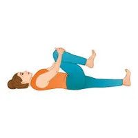
- Position: Supine lying position
- Raise one bent knee just far enough to allow you to use both hands to grab your lower leg. Just under the knee, interlace your fingers.
- If you are taking both up at the same time, clasp your wrists or interlace your fingers between your lower legs, just below your knees, just like in the single-legged variation.
- As much as you can, attempt to relax your low back, pelvis, and legs while you’re tugging. When performed passively, the knees-to-chest exercise more effectively engages the low back muscles.
- Continue on the opposite side. Hold duration is five to ten seconds.
- Repetition should be 10-15 times each session.
Piriformis stretch
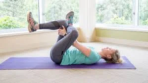
- Position: Supine lying position.
- Place one ankle across the thigh on the other side.
- Bring your knee up to your chest until your hips start to gently expand.
- Hold duration is five to ten seconds.
- Repetition should be 10-15 times each session.
Child pose
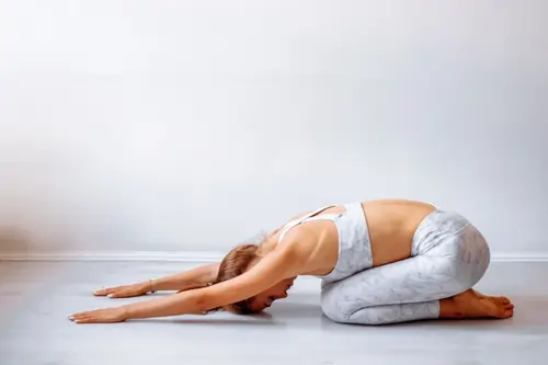
- Position: Sitting position.
- Kneel on the floor with your knees hip-width apart to do a child’s posture. After you’ve exhaled, drop your body between your knees. You can either relax your arms down the side of your torso or raise them over your head with your palms on the floor.
- Try a wide child’s position, which involves putting your knees farther apart and extending your arms forward between your legs, to feel an additional stretch in your hips and groin.
- Hold duration is five to ten seconds.
- Repetition should be 10-15 times each session.
Cat-Cow stretch
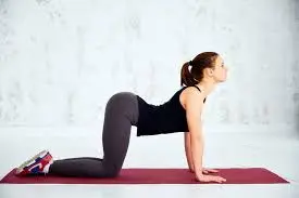
- Position: Quadruplicated position.
- With your spine in a neutral position, begin on your hands and knees.
- Ensure that your legs are hip-width apart and that your hands are precisely beneath your shoulders.
- Take a breath and raise your back in a cat position.
- After holding for a few seconds, exhale as you curve your back and tuck your chin like a cow.
- Hold duration is five to ten seconds.
- Repetition should be 10-15 times each session.
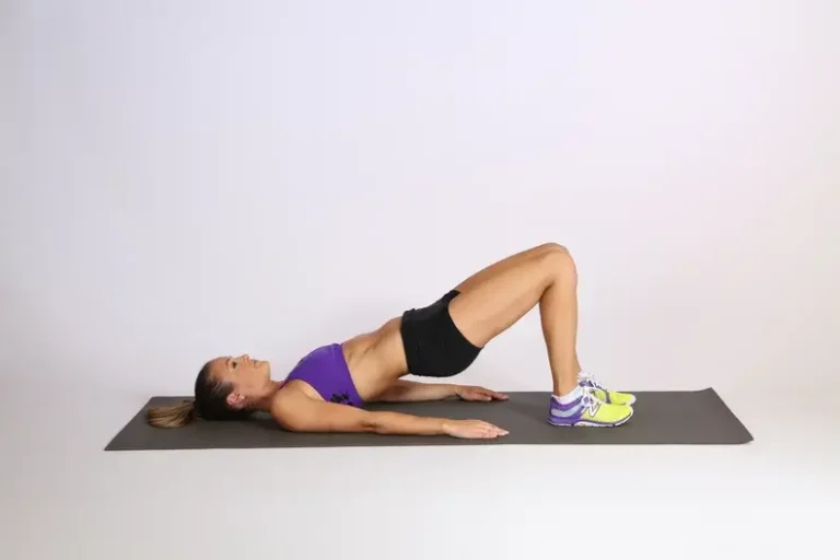
Bridging

- Position: Supine lying position.
- Keep your feet flat on the floor and your toes pointed straight forward as you slowly bend both knees.
- With the palms facing down, place the arms flat on the side of the body.
- Taking a breath, slowly lift your hips while using your buttocks and abdomen.
- To create a straight line from the shoulder to the knee, raise the hips as high as you can without experiencing any pain.
- Hold duration is five to ten seconds.
- Repetition should be 10-15 times each session.
Bird Dog pose

- Put your hands behind your shoulders and your knees beneath your hips.
- Use your abdominal muscles to keep your spine neutral.
- Your shoulder blades should be drawn together.
- Maintaining your shoulders and hips parallel to the floor, raise your left leg and right arm.
- To look down at the floor, drop your chin toward your chest, and lengthen the back of your neck.
- After a few seconds of holding this position, return to the beginning position.
- Elevate your right leg and left arm, and maintain this posture for a few seconds.
- Go back to where you were before. There is only one round.
- Hold duration is five to ten seconds.
- Repetition should be 10-15 times each session.
Variations of the bird dog exercise
- Weighted bird dog
- Pushup position
Side-Lying Clamshell

Clamshell steps:
- Position: side-lying position.
- With your knees bent in an “L” shape and your ankles stacked, lie on your side.
- Lift your top knee like you’re opening a clam shell while keeping your heels together.
- Return your knee to its starting position slowly.
- Hold duration is five to ten seconds.
- Repetition should be 10-15 times each session.
- Variations: Use a resistance band to strengthen your thigh muscles. Clam shelling will provide resistance with movement, and you can modify the resistance band.
Modified Plank

- Position: Prone position or side-lying position.
- Take a push-up stance to begin. Hold your position while lowering your knees to the floor.
- Hold duration is five to ten seconds.
- Repetition should be 10-15 times each session.
Modification:
- Side plank from knees: You should fold both knees and divide your body weight between your hands and knees, just like in the side plank exercise. This will provide some isometric workout for your obliques. Switch to the opposite side. Repeat the process.
- Hold duration is five to ten seconds.
- Repetition should be 10-15 times each session.
- Single-knee-side plank: Lower yourself from a sitting position so that your weight is evenly distributed between your hip and the side of your thigh closest to the floor. This leg’s slight bend will help to guarantee accurate and secure alignment.
- You have to think about the right alignment. Get your abs in shape. Keep one hand on your hip area and hold it straight.
- Try to add one or two seconds each time you practice, and concentrate on keeping the posture’s correct form. Hold duration is five to ten seconds.
- Repetition should be 10-15 times each session.
What more is there to do?
The following practices, in addition to routine stretches, may also lessen or avoid SI joint pain.
- Reducing extended sitting: Sitting for extended periods of time can make your back rigid and tight, which exacerbates SI joint discomfort. At least once every fifty to sixty minutes, try to get up and move about, stretch, or go for a little stroll.
- Altering your sitting posture: Maintaining your hips level with one another and your “sit bones” in touch with your chair is the ideal way to sit while suffering from SI joint pain. By doing this, the ligaments surrounding your SI joint may not be strained. Avoid crossing your legs and maintain a tiny distance between your knees. To preserve your back’s natural curvature, it may often be more comfortable for the SI joint if your hips are higher than your knees.
- Gluteal strengthening: Studies indicate that individuals with weak glutes and chronic SI joint discomfort may benefit from glute training.
- Alternating heat and cold: Try using alternating heat and ice for 15 to 20 minutes at a time to help control discomfort and increase blood flow. In the event that your discomfort doesn’t go away, think about physical therapy: To assist you in managing your SI joint discomfort, a physical therapist can create a personalized program.
Medicines
These might include the following, depending on the source of the pain:
- Painkillers. Ibuprofen (Advil, Motrin IB, and others) and naproxen sodium (Aleve) are examples of nonsteroidal anti-inflammatory pain medications that are available without a prescription. A doctor may recommend a more potent painkiller if they don’t offer adequate relief.
- Muscle relaxers. Cyclobenzaprine (Amrix) is one medication that may help lessen the muscular spasms that frequently accompany sacroiliitis.
- Biologicals. Numerous autoimmune diseases are treated using biologic medications. Ixekizumab (Taltz) and secukinumab (Cosentyx) are examples of interleukin-17 (IL-17) inhibitors. Etanercept (Enbrel), adalimumab (Humira), infliximab (Remicade), and golimumab (Simponi) are examples of tumor necrosis factor (TNF) inhibitors.
- DMARD medications reduce edema and discomfort while also reducing muscular inflammation. Some specifically target and inhibit the Janus kinase (JAK) enzyme. Tofacitinib (Xeljanz) and upadacitinib (Rinvoq) are examples of JAK inhibitors.
Procedures such as surgery
If pain has not been reduced by other means, a medical professional may recommend:
- The joint was shot. To lessen pain and swelling in the joint, corticosteroids might be used. Because the steroids might damage adjacent bones and tendons, you can only have a few joint injections annually.
- Radiation-induced denervation. The nerve that is generating the pain may be damaged or destroyed by radiofrequency radiation.
- Stimulation by electricity. Pain from sacroiliitis may be lessened by implanting an electrical stimulator in the lower spine.
- Fusion of joints. Fusing the two bones together with metal hardware can occasionally ease sacroiliitis discomfort, even though surgery is rarely used to treat the condition.
Special Test
There are several possible differential diagnoses for lower back pain.
Assessing gait, leg-length inequality, and the lower lumbar area are the first steps in the clinical assessment of a patient with sacroiliac joint dysfunction.
The Fortin finger test should be used to try to localize the SI joint during a physical examination. When the painful spot is located within 2 cm inferomedially to the posterior superior iliac spine, the test is deemed positive. To rule out further disorders, the hip joint and spine should be thoroughly examined in addition to the physical exam techniques mentioned above.
SI Joint Stress Tests
The Fortin finger test involves the patient using one finger to indicate the painful spot. If the pain location is within 1 cm of the PSIS, usually inferomedially, the result is positive.
The Gillette test is administered while the patient is standing. The patient flexes the opposing hip and knee into the chest while standing on one leg. One thumb is positioned behind the posterior superior iliac spine (PSIS) on the side of hip flexion, while the other thumb is in the midline at the S2 level to measure the SI joint’s range of motion. With hip flexion, the thumb under the PSIS often dips laterally and inferiorly. Reduced mobility in comparison to the usual side indicates restriction.
Other Special Test
- FABER Test
- Long sit test
- Sign of the buttocks
- Gaenslen’s Test
- Prone test
- Functional test
- Standing leg length test
Benefits
- Pain Reduction: By releasing tension in the surrounding muscles, gentle stretching and strengthening activities help relieve discomfort and pressure on the SI joint.
- Increased Stability: Pelvic stability, which is essential for shielding the SI joint from excessive movement or strain, is improved by strengthening the hip, glute, and core muscles.
- Increased Mobility: Specific motions improve range of motion and lessen stiffness by reestablishing hip and lower back flexibility.
- Improved Posture and Alignment: By promoting appropriate spinal and pelvic alignment, exercises can reduce compensatory tendencies and unequal loads, which frequently worsen SI joint discomfort.
- Decreased Inflammation: Regular exercise promotes tissue repair around the joint and helps wash out inflammatory chemicals by increasing circulation.
- Functional Independence: Exercise helps with everyday tasks like walking, sitting, and bending, which are frequently hampered by SI joint discomfort, by increasing strength and coordination.
- Prevention of Recurrence: By strengthening the musculoskeletal system’s resilience, a regular regimen reduces the likelihood of recurrent episodes or long-term instability.
Avoid Activity while experiencing SI joint pain.
- Tennis
- Partial or full crunches
- Golf
- Heavyweight lifting
- Football
- Basketball
- Prolonged sitting posture
FAQs
How can SI joint discomfort be resolved naturally?
Try alternating cold and heat therapy to ease inflammation and relax muscles, do low-impact, mild exercises and stretches to strengthen and support the joint, and adjust activities to prevent pain triggers like extended sitting if you have SI joint discomfort. Over-the-counter pain relievers can ease discomfort, and rest is essential. For a customized workout regimen for chronic pain, think about speaking with a physical therapist.
Does SI joint discomfort last a lifetime?
Because its length frequently varies depending on the reason, sacroiliitis is not necessarily permanent. Acute instances, such as those resulting from pregnancy or injury, frequently go away with rest and treatment. But if it’s connected to inflammatory arthritis, it may be persistent and need constant care.
What is the underlying reason behind SI joint pain?
Pain in the Sacroiliac Joint: Signs, Causes, and Management
Inflammation, trauma, or mechanical problems with the sacroiliac joints—which attach the sacrum to the pelvis—are the main causes of SI joint discomfort. Trauma, pregnancy, arthritis (including inflammatory and osteoarthritis), disparity in leg length, repeated stress, and sports injuries are common causes. The discomfort can also be caused by gout, spondylarthropathies, and the aftermath of spine surgery.
Which sleeping posture is ideal for those with SI joint pain?
Effective Sleep Techniques for Sacroiliac Joint Pain…
Try sleeping on your side with a cushion between your knees and ankles to preserve spinal and pelvic alignment, or on your back with a pillow beneath your knees, to relieve sacroiliac (SI) joint pain as you sleep. The SI joint may experience more stress and torque if you sleep on your stomach or in twisted, asymmetrical postures.
Does the SI joint show on lumbar MRI?
The lower lumbar spine, hips, pelvis, and the muscles and bones of the pelvic girdle are all visible during a standard scan procedure of the SI joints. As a result, incidental abnormalities that are not related to axSpA may be seen on SI joint MRIs; these findings may have clinical importance and should be reported.
What are the best exercises for reducing the pain of the sacroiliac joint?
You can perform a variety of exercises at home without the need for a trainer or any other equipment.
Knee to chest( single and double).
Clamshell.
Piriformis stretch.
Gluteus stretch.
Bridging.
Child pose.
Pelvic rotation.
Pillow Press.
Cat and camel stretch.
Bird dog stretch.
Side leg raise.
Prone leg raise.
Calf and Hamstring stretch.
References
- Cpt, D. R. M. C. (2023, June 23). Strengthening exercises for sacroiliac joint pain relief. Spine-health. https://www.spine-health.com/wellness/exercise/strengthening-exercises-sacroiliac-joint-pain-relief
- Yetman, D. (2025, August 1). Stretches and exercises to help ease SI (Sacroiliac) joint pain. Healthline. https://www.healthline.com/health/si-joint-stretches#bottom-line
- Sacroiliac Joint Pain Exercises & stretches for relief. (n.d.). https://www.romanomd.com/blog/sacroiliac-joint-pain-exercises–stretches-for-relief-20341.html
- Sacroiliitis – Diagnosis and treatment – Mayo Clinic. (n.d.). https://www.mayoclinic.org/diseases-conditions/sacroiliitis/diagnosis-treatment/drc-20350751
- Carr, J. (2024, April 8). Yoga for SI joint pain. PainTEQ | LINQ Procedure for SI Joint Dysfunction. https://www.painteq.com/yoga-for-si-joint-pain
- Cronkleton, E. (2023, October 27). How to perform the bird dog exercise and its core benefits. Healthline. https://www.healthline.com/health/bird-dog-exercise#alternative-exercises
- Cpt, A. A. (2024, August 6). Knees to chest stretch for low back muscles. Verywell Health. https://www.verywellhealth.com/knees-to-chest-exercise-296870
- Clinic, C. (2022, July 1). 7 Best stretches and exercises for Piriformis Syndrome. Cleveland Clinic. https://health.clevelandclinic.org/piriformis-syndrome-stretches-exercises
- TeachMeAnatomy. (2023, April 8). The sacroiliac Joint – Surfaces – ligaments – TeachMeAnatomy. https://teachmeanatomy.info/pelvis/bones/sacroiliac-joint/
- Wong, M., Sinkler, M. A., & Kiel, J. (2023, August 8). Anatomy, abdomen and pelvis, sacroiliac joint. StatPearls – NCBI Bookshelf. https://www.ncbi.nlm.nih.gov/books/NBK507801/
- Kiapour, A., Joukar, A., Elgafy, H., Erbulut, D. U., Agarwal, A. K., & Goel, V. K. (2019). Biomechanics of the sacroiliac joint: anatomy, function, biomechanics, sexual dimorphism, and causes of pain. The International Journal of Spine Surgery, 14(s1), S3–S13. https://doi.org/10.14444/6077
- 6 Ways to Ease Piriformis Syndrome Pain with Self-Massage and Stretches. (2021, March 10). Healthline. https://www.healthline.com/health/piriformis-massage#stretches